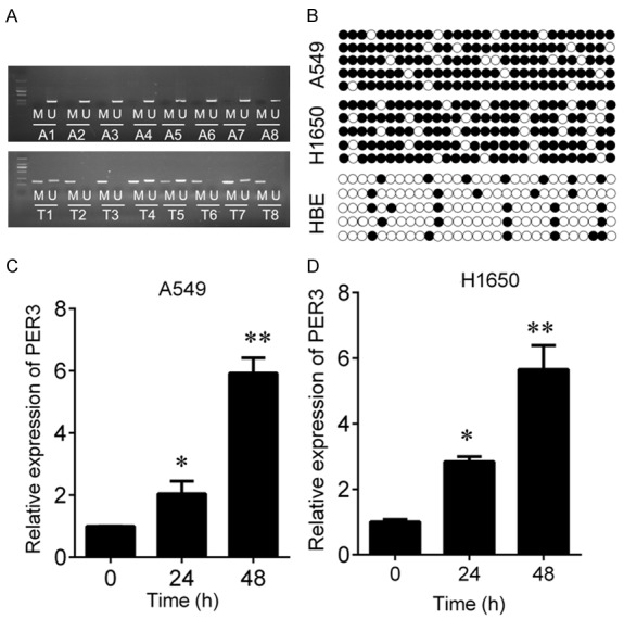
Figure 2.

PER3 is hypermethylated in NSCLC tissues and cells. A: MSP was used to measure the hypermethylation status of PER3 in NSCLC tissues and their matched adjacent tissues. M, methylation; U, unmethylation. B: BSP was used to measure the hypermethylation status of PER3 in HBE, A549 and H1650 cells. C, D: QPCR was used to measure the expression of PER3 in A549 and H1650 cells after 5-Aza treatment for 24 or 48 h. *P<0.05, **P<0.01.
