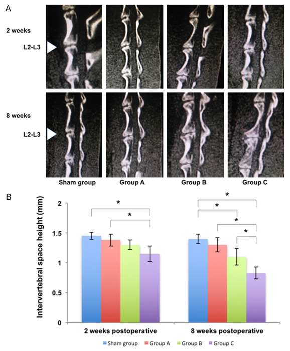
Figure 2.

CT sagittal reconstruction images and the middle height of punctured intervertebral space in different groups at 2 weeks and 8 weeks postoperatively. A. CT sagittal reconstruction images of the rabbit lumbar spine in different groups. The intervertebral space gradually narrowed in the L2-L3 IVD from sham group to Group C. There was calcification in Group C at 8 weeks postoperatively. B. The middle height of punctured intervertebral space in different groups. There was a significant difference between Group C and other groups at 8 weeks postoperatively (*P<0.05, unit: mm).
