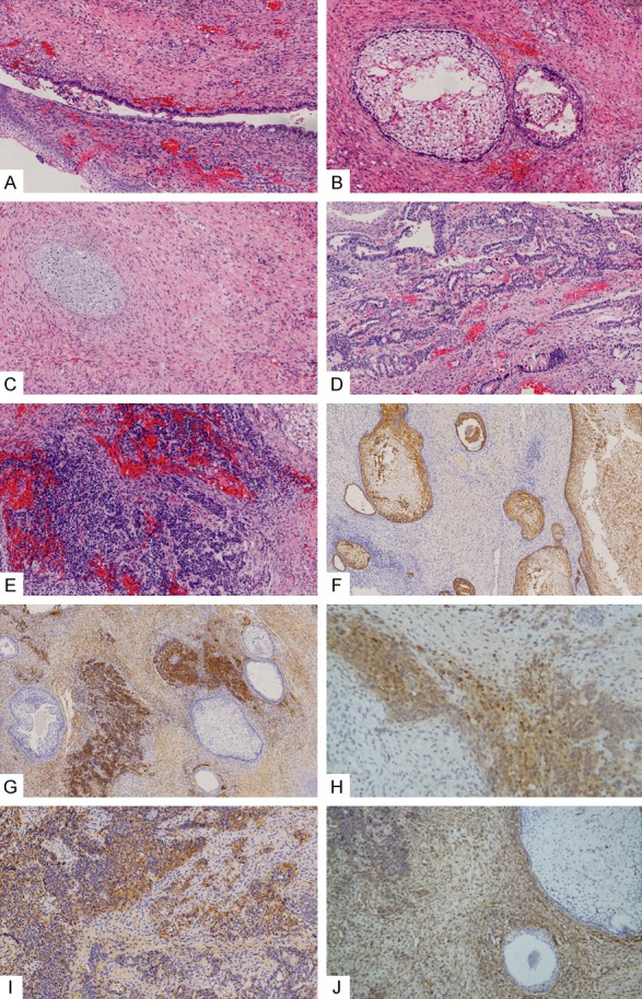
Figure 3.

Histopathological examination revealed the following signs: (A) pseudostratified ciliated columnar epithelium on the top of (B) immature squamous cell nests, chondrocytes (C) and adenocarcinoma structure (D) visible in some subepithelial regions, and massive dense proliferation of primitive small cells (E). (A-E: HE staining, ×200). Immunohistochemical staining revealed the following results: epithelial cells positive for epithelial CK (F), small cellular regions positive for CD56 (G) and NSE (H), some epithelial cells and small cells were positive for CD99 (I), and mesenchyma and small cells positive for Vim (J) (F and G: ×100; H-J: ×200).
