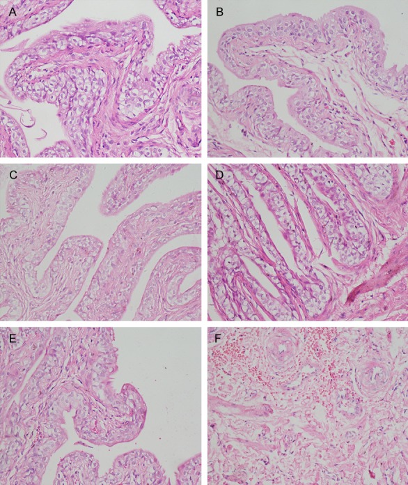
Figure 4.

Representative H&E staining (× 200). A: NS group at 1st week. No abnormality was observed; B: KET group at 1st week. Only a small amount of inflammatory cells infiltrated the bladder; C: NS group at 4th week. No abnormality was observed; D: KET group at 4th week. Most of the bladder epithelial showed necrosis and shedding, accompanied by microvascular congestion and bleeding; E: NS group at 12th week. No abnormality was observed; F: KET group at 12th week. The bladder epithelial cells appeared to include large amounts of red blood cells and with a large number of neutrophils infiltration.
