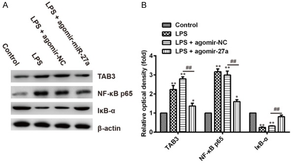
Figure 5.

miR-27a suppressed NF-κB pathway in lung tissues of LPS-induced ALI mice. A. The protein levels of TAB3, NF-κB p65 and IκB-α were assessed by Western blotting. β-actin were used as internal control. B. Quantification of protein expression was normalized to internal control using a densitometer. Data are shown as mean ± SD (n = 3). *P<0.05 **P<0.01 vs. Control group; ##P<0.01 vs. LPS + agomir-NC group.
