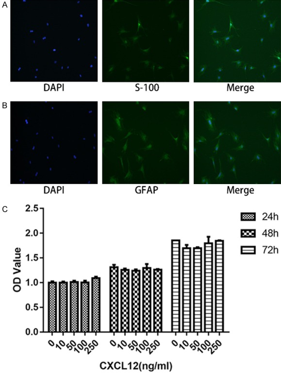
Figure 1.

Fluorescent immunocytochemistry of cultured Schwann cells stained by S-100 (A) and GFAP (B), with nuclei counterstained with DAPI. (C) Effects of different CXCL12 concentration on Schwann cell proliferation performed by CCK-8 assay. Data are presented as mean ± SD, n = 3. One-way ANOVA.
