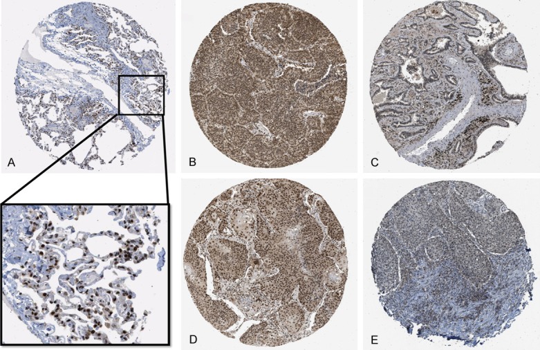
Figure 6.

IHC expression pattern of RBM5 in normal lung tissue and NSCLC from HPA database. A: Normal lung tissue expression of RBM5: the magnified area shows a strong expression pattern in the nuclei of pneumocytes. B: Representative strong expression of RBM5 IHC in lung adenocarcinoma. C: Representative weak expression in lung adenocarcinoma. D: Representative strong expression in lung squamous cell carcinoma. E: Representative weak expression in lung squamous cell carcinoma.
