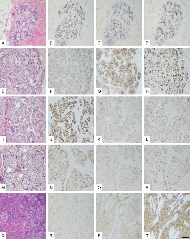
Figure 1.

Pathological characteristics of normal breast and IBC tissue. A-D. Normal breast tissue. A. Terminal ductal lobular structure (HE staining). B. ER expression in epithelial cells with strong and weak heterogeneity in the small lobule. C. p63 was positive in normal breast myoepithelial cells. D. CK5/6 showed a positive expression in normal breast glandular epithelium. E-H. HER2- overexpression breast cancer. E. Morphological features of infiltrating carcinoma (HE staining), cancer cells in invasive growth pattern. F. Cancer cells are ER negative. G. Immunohistochemical staining reveals HER2 positive cells (3+). H. High level of Ki67 expression with more than 50% nuclear positive cells. I-L. Luminal A type breast cancer. I. Morphological features of invasive carcinoma with no special type (HE staining). J. Immunohistochemically strong positive for ER with high level of expression (more than 90% nuclear positive cells), K. HER2 negative, L. Low expression of Ki67 with less than 5% nuclear positive cells. M-P. Luminal B type breast cancer. M. Morphological features of invasive carcinoma with no special type (HE staining), N. High expression for ER with more than 90% the nuclear positive cells. O. HER2-negative breast cancer. P. High level of Ki67 expression with more than 40% nuclear positive cells. Q-T. BLBC type breast cancer. Q. Morphological features of invasive carcinoma with no special type (HE staining). R. IHC, ER negative. S. IHC revealed EGFR positive cells (3+). T. IHC, Cytoplasmic positive (3+) for CK5/6. Scale bar, 100 um.
