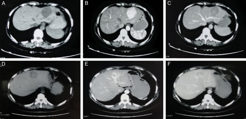
Figure 1.

Imaging characteristics of hepatic EAML. A 37-year-old woman with hepatic EAML in left lateral lobe of liver. (patient 1, A. Non-enhanced CT scan shows hypo-attenuating lesion in segment. B. Contrast-enhanced CT scan shows obviously enhanced lesion in the arterial phase. C. The lesion is hypo-attenuating in the portal venous phase). A 51-year-old woman with hepatic EAML in left lobe of liver. (patient 2, D. Non-enhanced CT scan shows hypo-attenuating lesion in segment. E. Contrast-enhanced CT scan shows of the homogeneous enhanced lesion with thickly distorted vessels in the arterial phase. F. The lesion shows partly prolonged hyper-attenuating in the portal venous phase).
