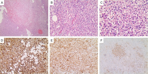
Figure 5.
Histopathology of hepatic EAML (patient 2). (A) Tumors mainly consisted of epithelioid cells that comprised approximately 95% of the total neoplastic mass (H&E stain, ×40). (B) The tumor was comprised of sheets of large polygonal cells with abundant granular eosinophilic cytoplasm (H&E stain, ×200). (C) Atypical epithelioid cells contain eosinophilic granular cytoplasm and pleomorphic nuclei (H&E stain, ×400). Immunohistochemical staining was positive for HMB-45 (D), Melan-A (E) and smooth muscle α-actin (F) (original magnification ×200).

