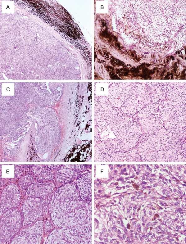
Figure 2.

(A) The primary mass showed a complete capsule with deep brown pigment around the mass (×40). (B) The recurrent mass showed a clear margin with the peripheral fat and muscle tissue (×100). (C) Capsular invasion was observed in the primary mass (×40). The nodules in the primary mass (D) (×100) and in the recurrent mass (E) (×200) were divided into packets, nests, or short cords by fibrous septa. (F) Tumor cells with mild atypia consisted of epithelioid cells or spindle cells with eosinophilic to clear cytoplasm, distinct round to oval nuclei, and prominent red nucleoli. Fine dust-like melanin pigment was occasionally observed in the cytoplasm (×400).
