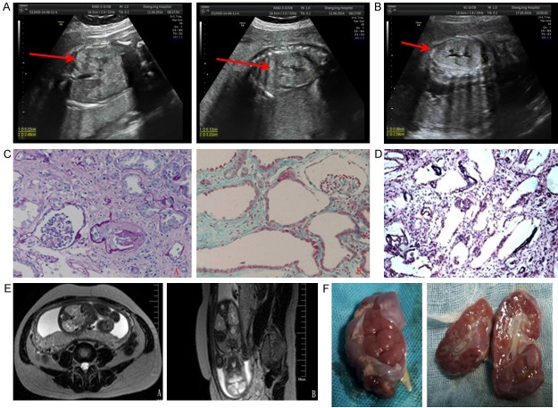
Figure 1.
(A) Fetal ultrasonography of the first proband showed bilateral kidney enlargement and enhanced echogenicity of the renal parenchyma (arrow). (B) Fetal ultrasonography of the second proband showed similar findings. (C) Renal biopsy of the second proband’s older brother showed massive cystic dilation of the proximal tubules with low-flat epithelium, epithelial cell exfoliation of partial tubules, as well as massive interstitial fibrosis (A: PAS 200 ×; B: Masson 200 ×). (D) Renal pathology of the first proband showed fusiform dilation of collecting ducts and distal renal tubules (light microscope, HE 100 ×). (E) Fetal magnetic resonance imaging of the second proband showed enlargement of both kidneys and mild dilation and deformation of the renal pelvis and calices (A: T2-weighted cross-sectional view, B: T2-weighted coronal view). (F) Gross examination of the fetal kidneys from the second proband, showing cystic enlargement.

