Figure 2.
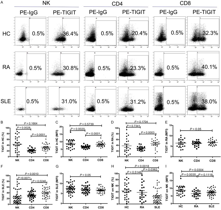
TIGIT expression on natural killer (NK) cells is decreased in patients with systemic lupus erythematosus (SLE). A. Representative dot plots of population gating and TIGIT expressing cells from healthy controls (HC), rheumatoid arthritis (RA) and SLE patients. Percentages of TIGIT expressing cells among NK, CD4+, CD8+ cells are shown. B. Summary data of the positive cell frequency in gated NK, CD4+, CD8+ cells from HC. C. Summary data of the mean fluorescence intensity (MFI) of TIGIT on NK, CD4+, CD8+ cells from HC. D. Summary data of the positive cell frequency in gated NK, CD4+, CD8+ cells from RA patients. E. Summary data of the mean fluorescence intensity (MFI) of TIGIT on NK, CD4+, CD8+ cells from RA patients. F. Summary data of the positive cell frequency in gated NK, CD4+, CD8+ cells from SLE patients. G. Summary data of the mean fluorescence intensity (MFI) of TIGIT on NK, CD4+, CD8+ cells from SLE patients. H. Summary data of the frequency of TIGIT-expressing NK cells from HC, RA and SLE patients. I. Summary data of the MFI of TIGIT on NK cells from HC, RA and SLE patients.

