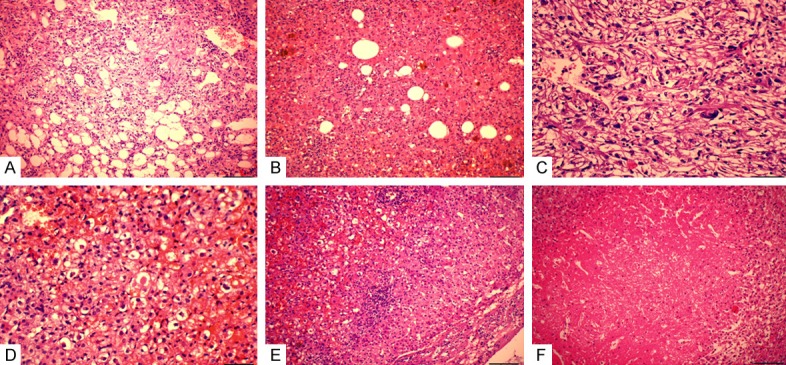
Figure 1.

Histology of PEComas. A. Epithelial smooth muscle cells were arranged in whorled and interlacing fascicles, surrounded by the tortuous vessels with the mature lipocytes being scattered. B. The epithelioid tumor cells of PEComas are polygonal or spheroidal, characterized by abundant cytoplasm that varied from eosinophilic granular to clear, with distinct cell border. C. Moderate cytologic atypia with enlarged vesicular nucleus and notable nucleolus, bizarre pleomorphic multinucleated giant cells were present. D. Extracellular hyaline globules were seen singly or in clusters. E. Epithelioid neoplastic cell invaded into the surrounding hepatic parenchyma without clear boundary. F. Local necrosis was present.
