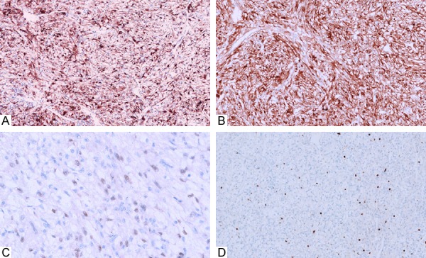
Figure 2.

Immunohistochemical study showed a strong and diffuse expression of HMB45 (A) in all the cases and Melan-A (B) in part of cases. Only 1 case showed the weak positive expression of TFE-3 (C) in the nucleus. The proliferation indexes were also low with less than 5% in all the cases (D).
