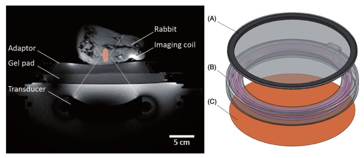
Figure 4.
Experimental set-up for mild hyperthermia in rabbit V × 2 tumors using a clinical MRI–HIFU system. Axial survey image of a rabbit on top of a water-filled animal adaptor. A waterproofed receive-only imaging coil is fitted around the lower leg. The bottom film of the animal adaptor is coupled to the window of the clinical HIFU system by a gel pad; the HIFU transducer is in the oil bath below. Overlays indicate the relative size of the ultrasound beam path (dashed) and treatment cell (shaded). Right: Rendering of the animal adaptor designed for the clinical HIFU system. The detachable lid (A) is a polyimide film glued to an acrylic ring. The cylindrical water bath (B) is a 3D-printed shell that holds a volume of degassed water, which is heated by water pumped through a coiled channel printed into the walls of the cylinder. Polyimide film (C) forms the base. Reproduced with permission from [108].

