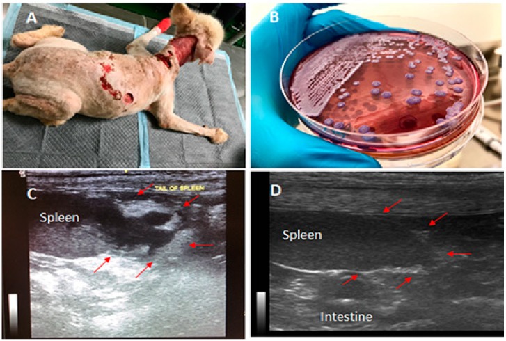
Figure 1.
Canine melioidosis: A, showing the massive wounds around the dog’s neck and back observed on the 1st day at Prince of Songkla University (PSU) Animal Hospital; B, Burkholderia pseudomallei colonies grown on Ashdown’s selective medium; C, the presence of a 2-cm abscess-like mass at the splenic tail confirmed by ultrasonography; and D, the recovery of the splenic tail after 10 weeks of the oral treatment. The arrows point to the areas of abscesses observed in C, which later disappeared after the oral treatment.

