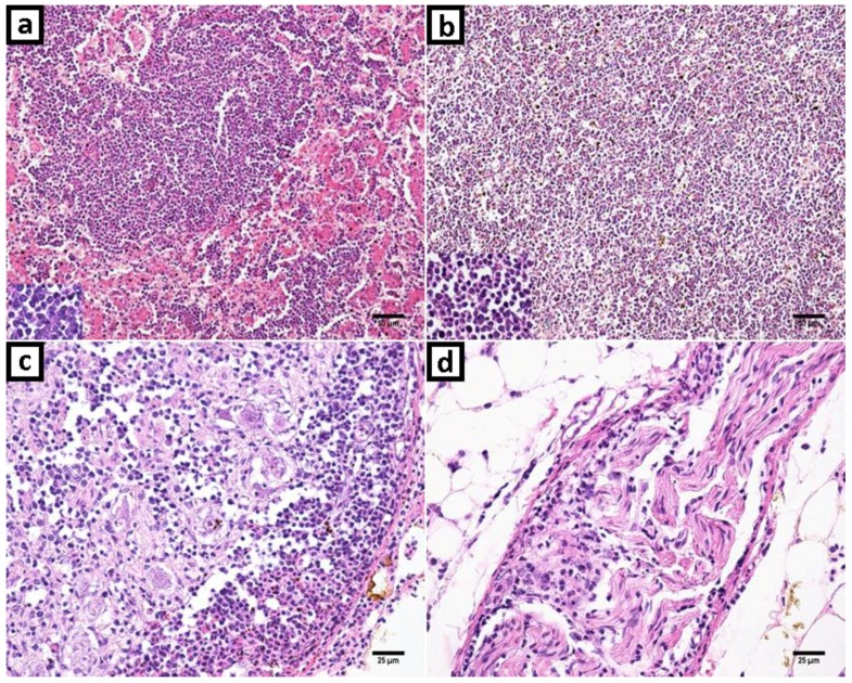
Figure 2.
Marek’s disease and reticuloendotheliosis in histopathology with hematoxylin and eosin (H&E) stanning. (a) Liver: Proliferation and infiltration of neoplastic small to medium size lymphocytes that replaced liver parenchyma. The neoplastic cells were uniform and small lymphocytes. (b) Spleen: Neoplastic lymphoid cells expanding and obliterating histologic structures; and with a highlight in detail of lymphocytes. (c) Ganglion: Neoplastic uniform small lymphocytes infiltration and hemorrhage foci. (d) Peripheric nerve. Increased cellularity due to small lymphocytes infiltration.

