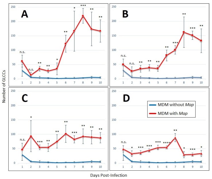
Figure 2.
GLCCs form in the presence of lymphocyte-specific signaling factors alone. Graphs of GLCC formation over ten-day experiment with MDMs cultured without non-adherent PBMCs, with conditioned medium from Map-exposed lymphocytes, after exposure to Map at an MOI of (A) 1:4, (B) 1:2, (C) 1:1, or (D) 2:1. The bars are standard deviation. * p < 0.05; ** p < 0.01; *** p < 0.001; **** p < 0.0001; n.s. = not significant by student’s t-test (n = 4).

