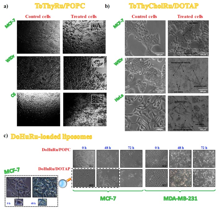
Figure 9.
Representative photomicrographs by phase-contrast light microscopy showing the cellular morphological changes and cytotoxic effects on cellular monolayers: untreated (left column in panels a and b) or treated for 48 h with ToThyRu/POPC (a) or ToThyCholRu/DOTAP (b) formulations. For ToThyRu/POPC nanosystems, MCF-7, WiDr and C6 cancer cell lines were examined (a); in turn, for ToThyCholRu/DOTAP nanocarriers, MCF-7, WiDr and HeLa cancer cell lines were analysed (b). Insets in (a) represent higher magnifications of injured cells following incubations with ToThyRu/POPC liposomes. (c) MCF-7 and MDA-MB-231 breast cancer cells treated for 48 and 72 h with ruthenium IC50 concentrations of DoHuRu-containing liposomes. The inset in (c) represents higher magnifications at 0 (left panel) and 48 h (right panel) DoHuRu/DOTAP treated MCF-7 cells, showing the formation of autophagic vacuoles detectable in cell cytoplasm. Figures are adapted from [77] (a), [79] (b) and [134] (c).

