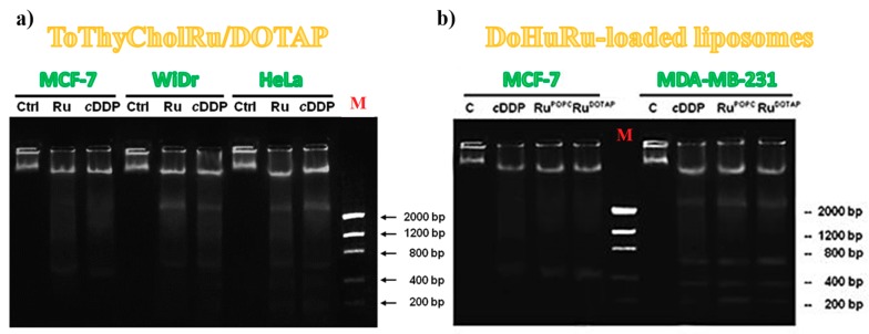
Figure 11.
1.5% agarose gels representing the DNA fragmentation assay on MCF-7, WiDr and HeLa cells, treated or not (Ctrl) for 48 h with IC50 doses of ToThyCholRu–DOTAP liposomes (indicated as Ru) (a) and MCF-7 and MDA-MB-321 cells treated or not (C) with IC50 concentrations of DoHuRu/POPC (RuPOPC) and DoHuRu/DOTAP (RuDOTAP) for 48 h (b). In both experiments, the selected cell lines were also treated with IC50 conc. of cisplatin (cDDP) used as a control. Lane M corresponds to the molecular weight markers. Figures are adapted from [79] (a) and [134] (b).

