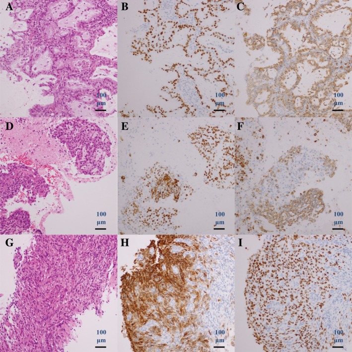
Figure 1.

Case 1. Histology of computed tomography‐guided percutaneous lung biopsy specimen before erlotinib therapy (A–C). (A) Haematoxylin and eosin, (B) TTF‐1 and CK5/6, and (C) p40 and napsin A staining. Histology of transbronchial lung biopsy specimen after erlotinib therapy, suggesting squamous cell transformation (D–F). (D) Haematoxylin and eosin, (E) TTF‐1 and CK5/6, and (F) p40 and napsin A staining. Histology of transbronchial lung biopsy specimen after combination chemotherapy (G–I). (G) Haematoxylin and eosin, (H) TTF‐1 and CK5/6, and (I) p40 and napsin A staining.
