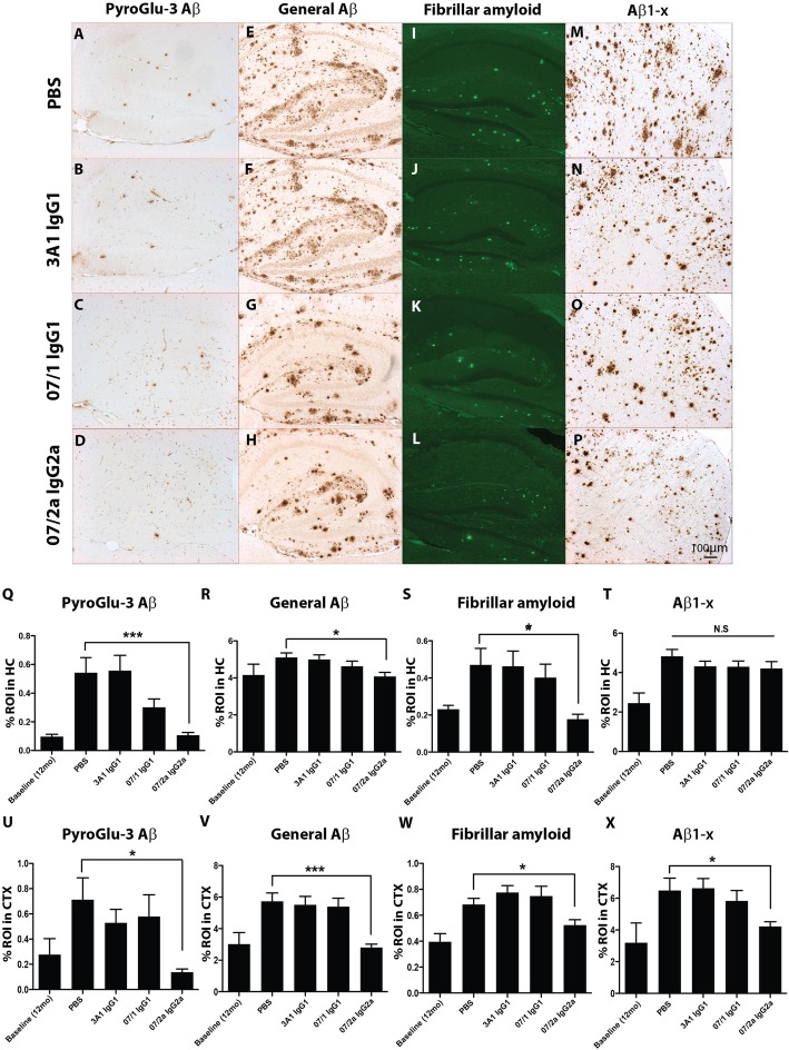
Fig. 2.
Lowering of multiple Aβ species in aged APPSWE/PS1ΔE9 Tg brains following 07/2a IgG2a mAb immunotherapy. Representative photomicrographs of hippocampus (HC) or cortex (CTX) from each treatment group of serial sections immunolabeled with a novel pGlu-3 Aβ IgG2b monoclonal antibody (a–d; HC), a general Aβ polyclonal antibody, R1282 (e–h; HC), stained with a marker for fibrillar amyloid, Thioflavin S (i–l; HC), and with a marker that specifically recognizes Aβ starting at Asp1, 82E1 (m–p; CTX). Quantitative image analysis on six stained sections at thee equidistant planes per marker demonstrated that there was a significant reduction of pGlu-3 Aβ (p < 0.001), general Aβ (p < 0.05), and fibrillar amyloid (p < 0.05) in the hippocampus of Tg mice immunized with 07/2a IgG2a mAb compared to Tg mice treated with PBS (q–t). Similarly, there was a significant reduction of pGlu-3 Aβ (p < 0.05), general Aβ, fibrillar amyloid, and Aβ1−x (p < 0.05) in the cortex of the Tg mice immunized with 07/2a IgG2a mAb compared to Tg mice treated with PBS (u–x). n = 13–15 per group. All data, excluding baseline, are expressed as the mean ± SEM. ANOVA with Neuman-Keuls post test: ***p < 0.001 and *p < 0.05 versus Tg PBS

