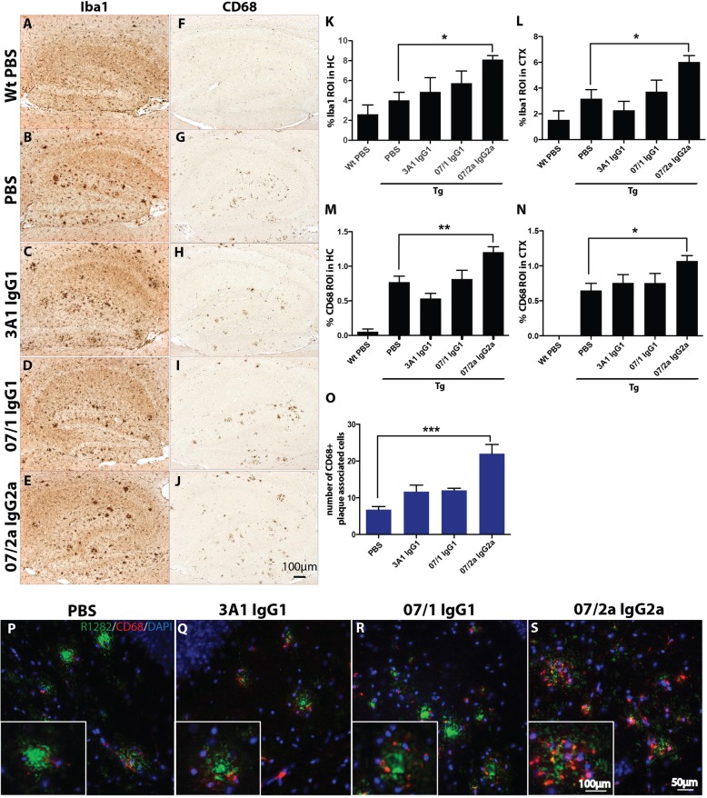
Fig. 4.
Immunohistochemical analysis of microglia and macrophage markers in the hippocampus and cortex of APPSWE/PS1ΔE9 Tg mice. Passive immunization, in aged APPSWE/PS1ΔE9 mice, resulted in a significant increase in plaque-associated microglia in mice treated with 07/2a IgG2a mAb. Representative photomicrographs of the hippocampus from each treatment group immunolabeled with microglial and macrophage marker, Iba1 (a–e), and activated microglial and macrophage marker, CD68 (f–j), showed an increased immunolabeling in immunized mice compared to PBS-treated Tg mice. Image analysis demonstrated that there was a significant increase in the percent area of Iba1-positive staining in the hippocampus (p < 0.05; k) and cortex (p < 0.05; l) of 07/2a IgG2a mAb-immunized mice compared to PBS-treated Tg mice. Analysis of the cortex demonstrated that there was a significant increase in CD68-positive immunolabeling in the hippocampus (p < 0.01; m) and cortex (p < 0.05; n) of 07/2a IgG2a mAb-immunized mice compared to PBS-treated Tg mice. A significant increase in plaque-associated CD68-positive cells was observed in Tg mice immunized with 07/2a IgG2a mAb (o, s). n = 13–15 per group. All data are expressed as the mean ± SEM. ANOVA with Neuman-Keuls post test: **p < 0.01 and *p < 0.05 versus Tg PBS

