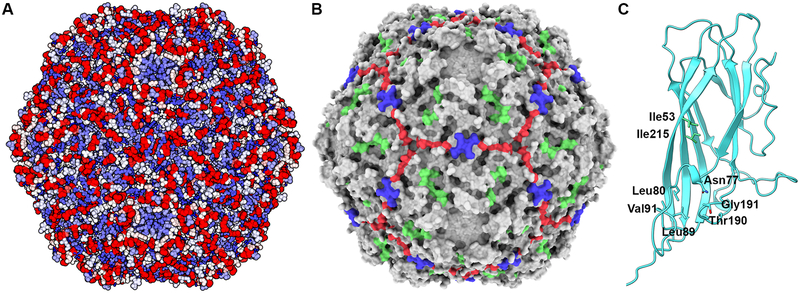
Figure 7. Sequence comparison of 1,278 PCV2 capsid protein entries plotted on the PCV2d atomic coordinates.
A) The sequence diversity and variation plotted on the PCV2d atomic coordinates -same coloring scheme as in Fig. 6. Image made with UCSF Chimera (Pettersen et al., 2004). B) Surface representation of the PCV2d atomic coordinates with the three conserved patches colored in green (Tyr55, Thr56, Asp70, Met71, Arg73, Asp127), blue (Pro82, Gly83, Gly85) and red (Asp168, Thr170, Gln188, Thr189). Image made with UCSF ChimeraX and colored using flat lighting (Goddard et al., 2018). C) Ribbon cartoon of a PCV2d subunit. Amino acids in stick are evolutionary coupled together, as determined using the plmc. MATLAB 2019, and EVzoom programs. Figures generated using UCSF Chimera (Pettersen et al., 2004).

