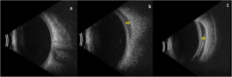
Fig. 8.
a-c Ultrasonographyic view of WJ-MSC implantation into the deep subretinal space within the extraocular muscle conus; a: before the application (Table 1, patient no. 1), b: injection via 25 G sharp-tip needle (Table 1, patient no. 1), c: placement via 20 G curved subtenon canulla with pre-placed suture to prevent the leakage (Table 1, patient no. 4)

