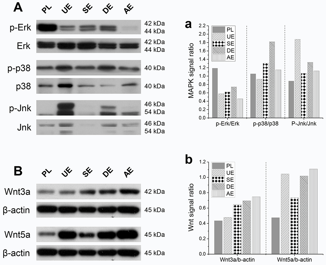
Fig. 5.

Potential mechanisms involved in the rejuvenation of SDSCs after expansion on dECMs [SECM (SE), DECM (DE), AECM (AE), and UECM (UE)]. Western blot was used to detect the mitogen-activated protein kinase (MAPK) signals (A), including Erk, p38, and Jnk, and the Wnt signals (B), including Wnt3a and Wnt5a. The intensity of immunoblotting bands was measured using ImageJ software and MAPK signals were presented as a ratio of phosphorylated protein to non-phosphorylated protein (a). The expression of Wnt signals was quantitated and normalized with β-actin (b). This experiment was repeated three times.
