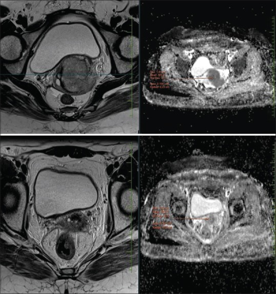
Figure 1.

Case of Ca cervix. Top row shows pre chemo radiation mass seen in uterine cervix (mean ADC values marked). Bottom row shows post treatment fibrotic changes with ill-defined enhancing thickening predominantly involving the anterior lip of cervix. ADC values are not representative of residual disease. Histopathological correlation confirmed the above findings
