Figure 6 (A-C).

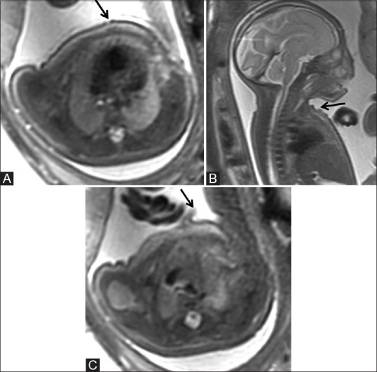
Axial (A) and sagittal (B) T2-weighted images of the fetus showing irregular skin surface with focal areas of discontinuities (arrow) along the anterior chest wall (C) and small focal skin flap, possibly due to exfoliation (arrow)

Axial (A) and sagittal (B) T2-weighted images of the fetus showing irregular skin surface with focal areas of discontinuities (arrow) along the anterior chest wall (C) and small focal skin flap, possibly due to exfoliation (arrow)