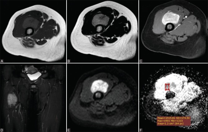
Figure 2 (A-F).
Paraganglioma in a 20-year-old woman (A) Axial T1WI and (B) Axial T2WI and (C) Postcontrast Axial THRIVE WI (D) Coronal STIR WIs showed a well-defined soft tissue mass is seen involving the anterior thigh compartment at a deep peri-osseous location eliciting isointense to low signal on T1 and heterogeneous isointense and high T2/STIR signal. with intense homogeneous enhancement in postcontrast images. Corresponding DWI (E) and ADC maps (F) showed high signal in DWI and low ADC signal with a ADCmean value = 1.55 × 10⁻³ mm2/s

