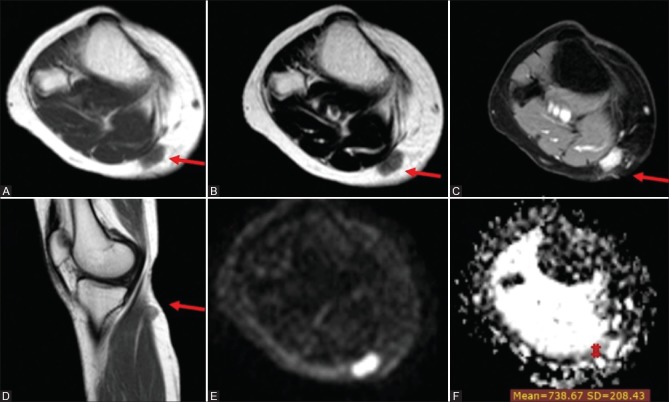
Figure 3 (A-F).
Unclassified soft tissue sarcoma in a 30-year-old woman (A) Axial T1WI (B) Axial T2WI and (C) postcontrast axial Thrive WI and (D) postcontrast sagittal T1 WIs showing well-defined superficial soft tissue nodule seen involving the posteromedial aspect of the upper leg at a subcutaneous location inseparable from the fascia of the medial head of gastrocnemius muscle. It elicits an isointense signal to muscle on T1 WI, isointense to a high signal on T2 WIs with intense homogeneous enhancement on postcontrast series. Corresponding DWI (E) and ADC map (F) showed high signal in DWI and iso to low signal on ADC map with an ADCmean value = 0.74 × 10⁻³ mm2/s

