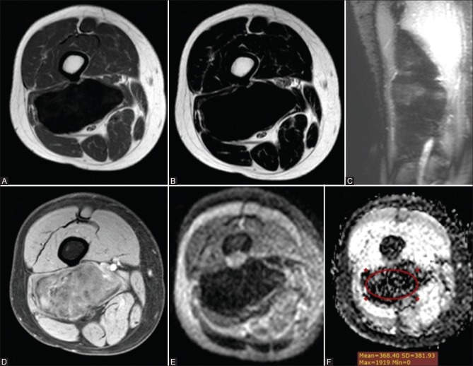
Figure 4 (A-F).
Fibromatosis in a 35-year-old man (A) axial T1WI (B) axial T2 WI and (C) coronal STIR WIs showing a well-circumscribed deep soft tissue mass in the posterior muscular compartment of the right thigh along the biceps femoris muscle abutting the lateral aspect of the lower femoral vessels eliciting marked hypointense signal on T1, T2 and STIR WIs. (D) Postcontrast axial THRIVE WIs showed mild heterogeneous enhancement. Corresponding DWI (E) and ADC map (F) showed low signal in DWI and ADC map with a mean ADC value = 0.37 × 10⁻³ mm2/s

