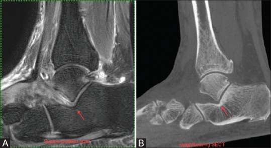
Figure 13 (A and B).

Same patient as Figure 12: Sagittal MRI in neutral position (A) do not show apposition between the bony surfaces with intervening edematous soft tissue seen similar to that seen in cases with extra-articular talocalcaneal impingement (arrow), (B) The sagittal reformatted images of the WBCT shows persistence of the distance between the bony surfaces without any collapse or direct contact between the bony surfaces as seen in the cases with extra-articular talocalcaneal impingement
