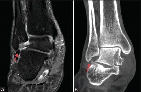
Figure 14 (A and B).

Subfibular impingement in a patient with flat foot (A) coronal MRI in neutral position show mildly reduced distance between the tip of the lateral malleolus and the lateral calcaneal process. (arrow), (B) coronal reformatted images of the WBCT shows further reduction of the distance with almost apposition of the bony surfaces (arrow)
