Figure 5 (A and B).

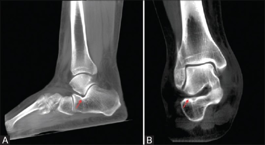
Normal appearance of the extra-articular talocalcaneal space on weight-bearing scans. Note the normal gap between the apposing bony surfaces on the sagittal (A) as well as coronal (B) reformatted thick slab images (arrows)

Normal appearance of the extra-articular talocalcaneal space on weight-bearing scans. Note the normal gap between the apposing bony surfaces on the sagittal (A) as well as coronal (B) reformatted thick slab images (arrows)