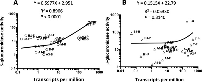
Fig. 10.
a and b Linear regression analysis of plots of TPM versus β-glucuronidase activity for each promoter-gusA fusion under the indicated conditions in liquid-grown cells (a) and plate grown cells (b). The media used were, B, BHI; P, PGY; F, FABG. Promoter fusion were to pilA1 (A1), pilA2 (A2), pilA3 (A3), pilB1 (B1), pilD (D), pilM (M), pilT (T). The line formulas, R2 and P values are shown for each data set. Note both panels are in log scales on each axis. The P values were calculated to determine if the slope is significantly non-zero

