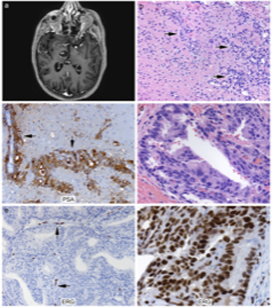
Figure 1.

A, a representative MR Image of a right thalamic metastasis. Figure 1B (H&E, 200x), in the thalamic metastasis example, there is extension of metastasis along Virchow-Robin spaces. Figure 1B (H&E, 200x) thalamic metastasis (arrows indicate tumor cells). Figure 1C (200x), the thalamic metastasis showed diffuse strong cytoplasmic immunoreactivity for prostate specific antigen (arrows). Figure 1D (H&E, 400x), most dural and parenchymal cases showed prototypical prostatic carcinoma histological features with cribriform gland formation. Figure 1E (ERG, 200x), a representative case of ERG negative dural-based tumor in contrast to the ERG-positive endothelial cells (arrows). Figure 1F, a representative case of ERG positive tumor with typical nuclear positivity.
