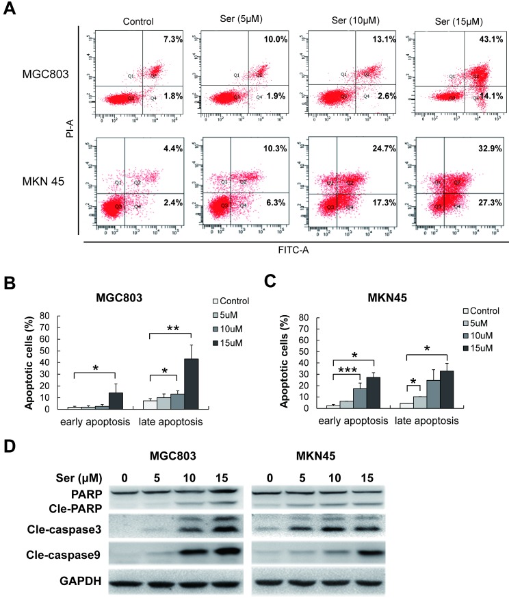
Figure 3.
Sertindole induced cell apoptosis in GC cells. (A) MGC803 and MKN45 cells were treated with gradient concentrations of sertindole for 24 h. Then, the cells were stained with PI at 37 °C for 30 min and measured by flow cytometry after collection. (B) Quantitative analysis of apoptotic cells. The percentage of cells in different phases of cell apoptosis was represented by a bar diagram. Data are presented as the mean ± SD of three independent experiments. (C) Effects of sertindole on the expression of apoptotic-associated proteins. (D) MGC803 and MKN45 GC cells were treated with 0, 5, 10 and 15 μM of sertindole for 24 h, and cell lysates were subjected to western blot analysis with cleaved PARP, caspase-3 and caspase-9 antibodies.

