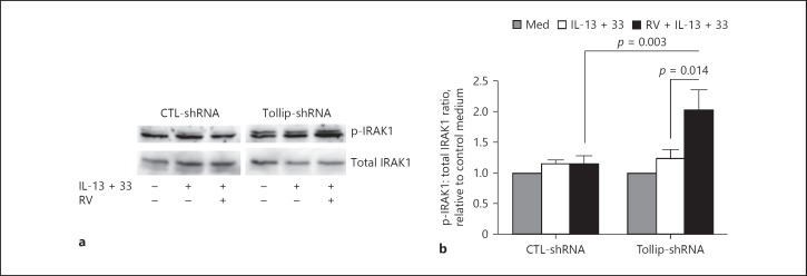
Fig. 3.
IRAK1 activation in Tollip-deficient (Tollip-shRNA) and Tollip-sufficient (CTL-shRNA) primary human tracheobronchial epithelial cells. a Representative Western blot showing p-IRAK1 and total IRAK1 expression levels. RV, rhinovirus. b Densitometric analysis of p-IRAK1 expression that was normalized to total IRAK1 expression. Results are expressed as fold change relative to unstimulated control (Med). Data are means ± SEM of n = 4 donors.

