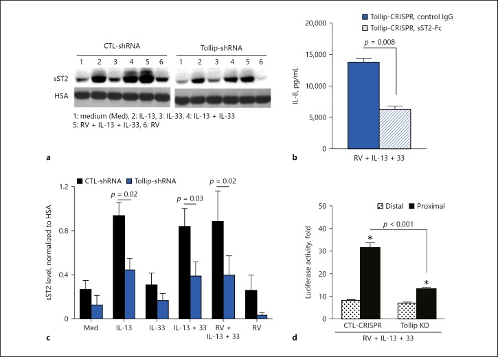
Fig. 6.
a Representative Western blot showing soluble ST2 (sST2) expression levels in culture supernatants of Tollip-deficient (Tollip-shRNA) and Tollip-sufficient (CTL-shRNA) primary human tracheobronchial epithelial (HTBE) cells. HSA, human serum albumin. b Effect of exogenous sST2 (sST2-Fc) on IL-8 production by Tollip knockout (KO) HTBE cells. sST2-Fc (1 µg/mL) was added to the culture 2 h before infection. IL-8 was measured 24 h after the infection. c Densitometric analysis of sST2 levels in culture supernatants of Tollip-deficient (Tollip-shRNA) and Tollip-sufficient (CTL-shRNA) HTBE cells (means ± SEM of n = 4 donors). d Differential usage of the ST2 promoter in control and Tollip KO HTBE cells after rhinovirus (RV) infection in the presence of IL-13 and IL-33. Data are means ± SEM of n = 4 replicates for each condition. * p < 0.05, compared to CTL-shRNA.

