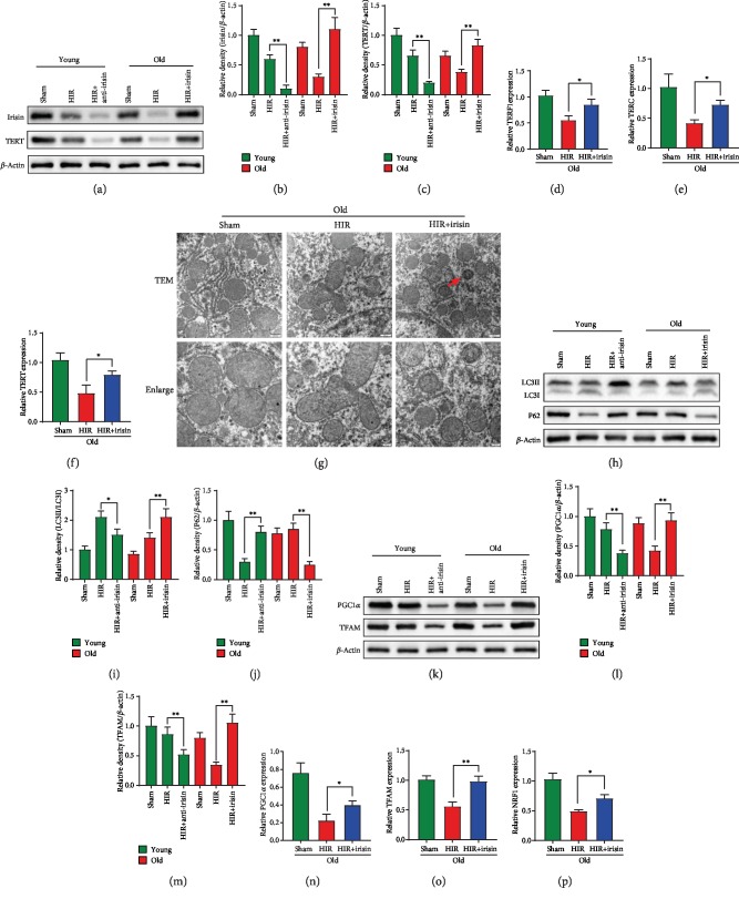
Figure 3.
Irisin improved telomerase activity, autophagy, and mitochondrial function in old rats after hepatic IR. Partial (70%) liver arterial/portal venous blood was interrupted for 40 minutes in 3-month- and 22-month-old rats. Irisin was administrated in old rats (iv. 250 μg/kg, single dose) at the beginning of reperfusion. Irisin-neutralizing antibody was administrated in young rats (iv. 50 μg/kg, single dose) at 24 h before hepatic IR. Liver samples were harvested at 24 h after reperfusion. (a–c) Western blot analysis of liver irisin and TERT expression. (d–f) qPCR analysis of liver TERF1, TERC, and TERT expression in old rats. (g) TEM analysis. The red arrow indicates autophagosomes. (h–j) Western blot analysis of liver LC3B and P62 expression. (k–m) Western blot analysis of liver PGC1α and TFAM expression. (n–p) qPCR analysis of liver PGC1α, TFAM, and NRF1 expression; n = 6; mean ± SEM; ∗P < 0.05 and ∗∗P < 0.01.

