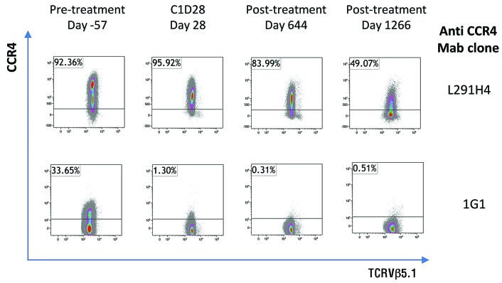
Figure 2.
Flow cytometric staining of CCR4 expression on malignant cells. PBMC from patient A1 were stained as described in Rowan et al.14: with a viability stain and antibodies specific for CD3, CD4, CD8, TCRVβ5.1 and with two different anti-CCR4 mAb clones. Cells were gated on live, single, CD3+CD8−TCRVβ5.1+ lymphocytes based on fluorescence minus one controls.

