Figure 1.
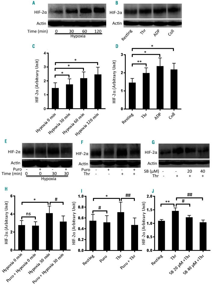
Enhanced expression of HIF-2α in human platelets under hypoxia and upon stimulation with physiological agonists. (A, B) Western blot analysis showing expression of HIF-2α in platelets exposed to either hypoxia (1% O2, 5% CO2, and 94% N2) for the indicated times (A) or agonists (thrombin, Thr, 1 U/mL; ADP, 10 μM; collagen, Coll, 10 μg/mL) under non-stirring condition for 10 min at 37°C (B). (C, D) Corresponding densitometric analyses of HIF-2α normalized to β-actin (n≥3). (E, F) Expression of HIF-2α in platelets pretreated or not with puromycin (Puro, 10 mM) and then exposed to hypoxia (E) or thrombin (F). (H, I) Corresponding densitometric analyses of HIF-2α normalized to β-actin ((n≥4). (G) HIF-2α expression of platelets pretreated with SB202190 (SB, 20 μM and 40 μM). (J) Corresponding densitometric analysis of HIF-2α normalized to β-actin (n=6). Data are represented as the mean ± standard error of mean of at least three different experiments. *P<0.05; **P<0.01; #P<0.05; ##P<0.01, analyzed by the Student t test.

