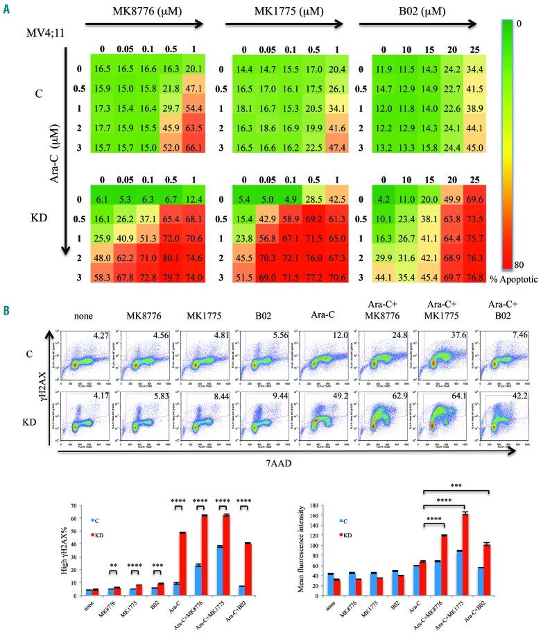
Figure 4.
Suppression of DOCK2 expression rendered MV4;11 cells more sensitive to MK8776, MK1775 and B02. (A) Compared to control cells, DOCK2 knokcdown (KD) MV4;11 cells exhibited increased percentages of apoptotic cells upon treatment with MK8776, MK1775 and B02, both alone and in the presence of ara-C. Cells were treated for 48 h. The assays were performed in triplicate. (B) Compared to control cells, a higher percentage of DOCK2 KD MV4;11 cells harbored elevated DNA damage (as indicated by an elevated gH2AX signal) upon treatment with ara-C (2 μM), as well as with MK8776 (0.1 μM), MK1775 (0.1 μM), and B02 (20 μM), both alone and in combination with ara-C. Treatment with MK8776, MK1775 and B02 in combination with ara-C resulted in an increased mean gH2AX signal in DOCK2 KD MV4;11 cells compared to cells treated with ara-C alone. Cells were treated for 16 h. *P<0.05; **P<0.01; ***P<0.001; ****P<0.0001. C: cells expressing control short hairpin (sh)RNA; KD: cells expressing an shRNA against DOCK2.

