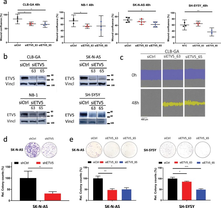
Figure 2.
ETV5 is required for cell migration and colony formation of neuroblastoma cells in vitro. (a) Wound confluence at 48 h after wound making on IncuCyte® of four different neuroblastoma cell lines after ETV5 knockdown (siETV5_63 and siETV5_65) compared to control vector (siCtrl). (nCLB-GA, NB-1, SH-SY5Y = 3; nSK-N-AS = 5; mean with error bars representing SD after error propagation). (b) Western blot analysis for ETV5 at 48 h after ETV5 knockdown (siETV5_63 and siETV5_65) in four different neuroblastoma cell lines. (vertical line indicates cropped image, full-length images are presented in Supplementary Fig. S7b). (c) Phase contrast imaging of the scratch wound on IncuCyte® for CLB-GA at start (0 h) and after 48 h of ETV5 knockdown (siETV5_63 and siETV5_65) compared to control vector (siCtrl). Blue represents the initial scratch wound, yellow represents the scratch wound masking. (d,e) Colony formation analysis of two different NB cell lines after ETV5 knockdown with shETV5 compared to control vector (shCtrl SHC002; left panel) and after ETV5 knockdown with siETV5_63 and siETV5_65 compared to control vector (siCtrl; middle and right panel). The upper panel shows the cristal violet staining, the lower panel shows the relative colony counts. (nshRNA = 2; nsiRNA, SK-N-AS = 4; nsiRNA, SH-SY5Y = 3; mean with error bars representing SD after error propagation) (*p < 0.05; **p < 0.01; ***p < 0.001).

