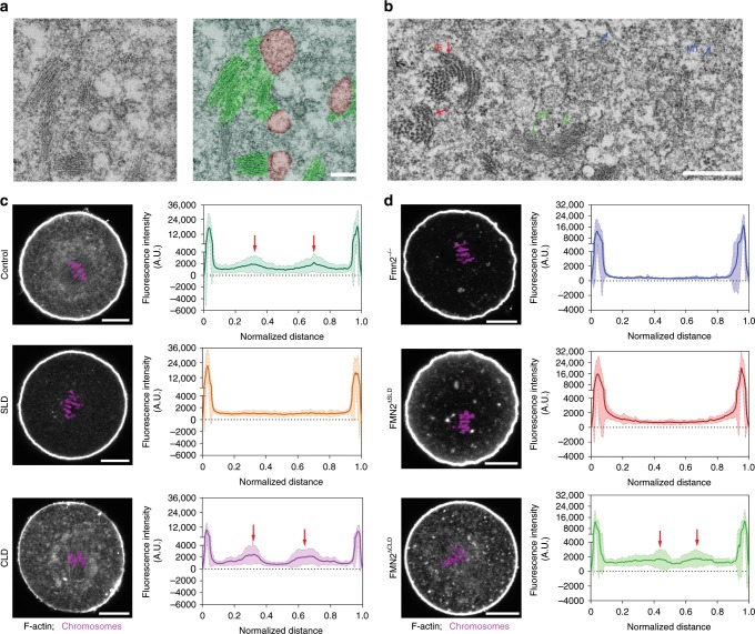
Fig. 2. Spindle peripheral FMN2 is required for actin polymerization surrounding the spindle.
a Representative EM images of MI oocytes show that short bundles of actin filaments form comet tail-like end-on association with the surface of ER-like vesicles. Red, ER-like vesicles. Green, F-actin bundles. Representative results, n = 3. Scale bar, 0.2 µm. b Representative EM images of MI oocytes show different appearance and thickness of microfilament (MF, green arrow), intermediate filament (IF, red arrow) or microtubule (MT, blue arrow). Representative results, n = 3. Scale bar, 0.5 µm. c Representative images of rhodamine phalloidin staining of F-actin after SLD or CLD expression in wild-type oocytes. The accumulation of F-actin surrounding the spindle was disrupted in oocytes expressing SLD but not CLD. The curves show the average of fluorescence intensity trace of actin along a thick line crossing the mid-zone of the MI spindle and the shades show the SD of the central lines (see Supplementary Fig. 1c). Red arrows point to actin intensity peaks around the spindle. n = 21 (control), n = 13 (CLD), n = 21 (SLD). Scale bar, 20 µm. d Representative images of phalloidin staining in Fmn2−/− oocytes with FMN2∆SLD or FMN2∆CLD expression. FMN2∆CLD rescued the spindle peripheral F-actin accumulation in Fmn2−/− oocytes. Line traces of average fluorescence intensity of actin and SD (shade) are shown as in c. n = 19 (Fmn2−/−), n = 15 (FMN2∆CLD), n = 17 (FMN2∆SLD). Scale bar, 20 µm.

