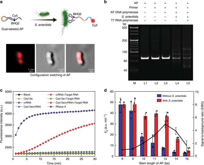
Fig. 2. Analysis of APC-Cas.
a Illustration and representative laser scanning confocal microscope (LSCM) images of dual-labeled AP binding to the S. Enteritidis. b Electrophoretic analysis of the feasibility of APC-Cas for S. Enteritidis detection. M: DNA marker, L1: AP; L2: AP + primer; L3: AP + primer + KF DNA polymerase; L4: AP + primer + KF DNA polymerase + S. Enteritidis; L5: AP + primer + KF DNA polymerase + S. Enteritidis + T7 RNA polymerase. ‘ + ’ means presence, ‘-’ means absence. c Fluorescence measurement of LbuCas13a activity. RNase A was used as positive control for the degradation of RNA reporter probe. d Comparison of seven APs with varied stem-length. Data represent mean ± s.d., n = 3, three technical replicates.

