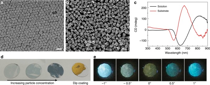
Fig. 6. Morphology and optical characteristic of the substrates coated with 432 helicoid IV.
a, b Large-area SEM image of the substrate coated with nanoparticles by the dip-coating method. Two different types of 432 helicoid IV, made with 0.1 mM (a) and 0.2 mM (b) of Cys concentration, were used for coating. A monolayer or bilayer of assembled nanoparticles was uniformly generated on the substrate. c CD spectra of nanoparticles in the solution phase (black) and coated on the substrate (red). The coupling of assembled nanoparticles in the film-type results in a significant change in spectral features compared with the results of the nanoparticle solution. d Photograph of 432 helicoid IV assembled substrates with different concentrations of nanoparticles. For the dip-coating sample, which has the highest packing density, the gold reflection color is shown. e Polarization-resolved transmission image of a substrate coated with nanoparticles. The angle of the analyzer was changed from −1° to 1°. See the Methods section for coating procedures and polarization experiments. Scale bars, 200 nm.

