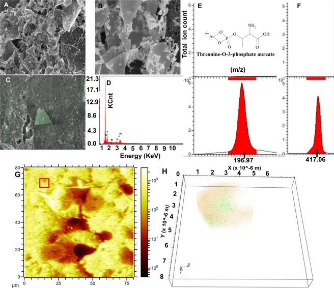
Figure 6.
SEM and ToF-SIMS analysis of de novo biomineralized nanostructures at 2 mM Au ions: delta and rhombus shaped particles. (A–D) SEM and EDX chemical mapping to confirm anisotropic rhombus/delta shape Au nanoplate formation (Scale bar 1 micron). (E,F) The SIMS signals from Au+ (E) and threonine-O-3-phosphate aureate (F). (G) Reconstructed ToF-SIMS ion image from the A549 cell surface demonstrating spherical Au particles. (H) The 3D reconstruction and overlay of Au+ signal (orange) and threonine-O-3-phosphate aureate (green) from red square ROI from (G).

