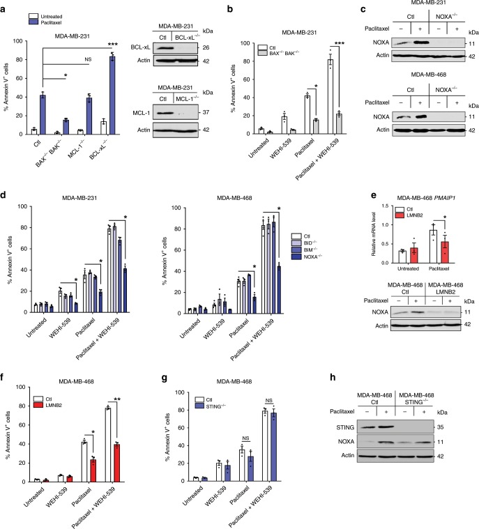
Fig. 6. NOXA is induced by micronuclei envelope collapse-dependent but STING-independent signals.
a Annexin V assay in control, BCL-xL or MCL-1 KO MDA-MB-231 cells treated or not with paclitaxel for 24 h, washed out, incubated for 48 h (left panel). Analysis of BCL-xL and MCL-1 expression in untreated cells was realized by immunoblot (right panel). b Same analysis as in a in control or double KO BAX−/− BAK−/− MDA-MB-231 cells treated with paclitaxel for 24 h or not, washed out for 48 h then treated with WEHI-539 for 24 h or not (c). Immunoblot analysis in control or NOXA−/− breast cancer cells 48 h after a 24 h-paclitaxel treatment. d Same experiment as (b) using control, BID−/−, BIM−/−, or NOXA−/− MDA-MB-231 or MDA-MB-468 cell lines. e PMAIP1 qPCR (upper panel) and NOXA immunoblot (lower panel) in control or LMNB2 overexpressing MDA-MB-468 cells 48 h after a 24 h-paclitaxel treatment. f Same experiment as (b) using control or LMNB2 overexpressing MDA-MB-468 cells. g Same experiment as (b) using control or STING−/− MDA-MB-468 cells and indicated treatments. h NOXA immunoblot analysis in control and STING−/−MDA-MB-468 cells harvested 48 h after a 24 h-paclitaxel treatment. Data were collected from n = 3 independent experiments. Error bars indicate mean +/− SEM; Two-sided paired t-test. The symbols correspond to a p-value inferior to *0.05, **0.01, and ***0.001. NS: not significant.

